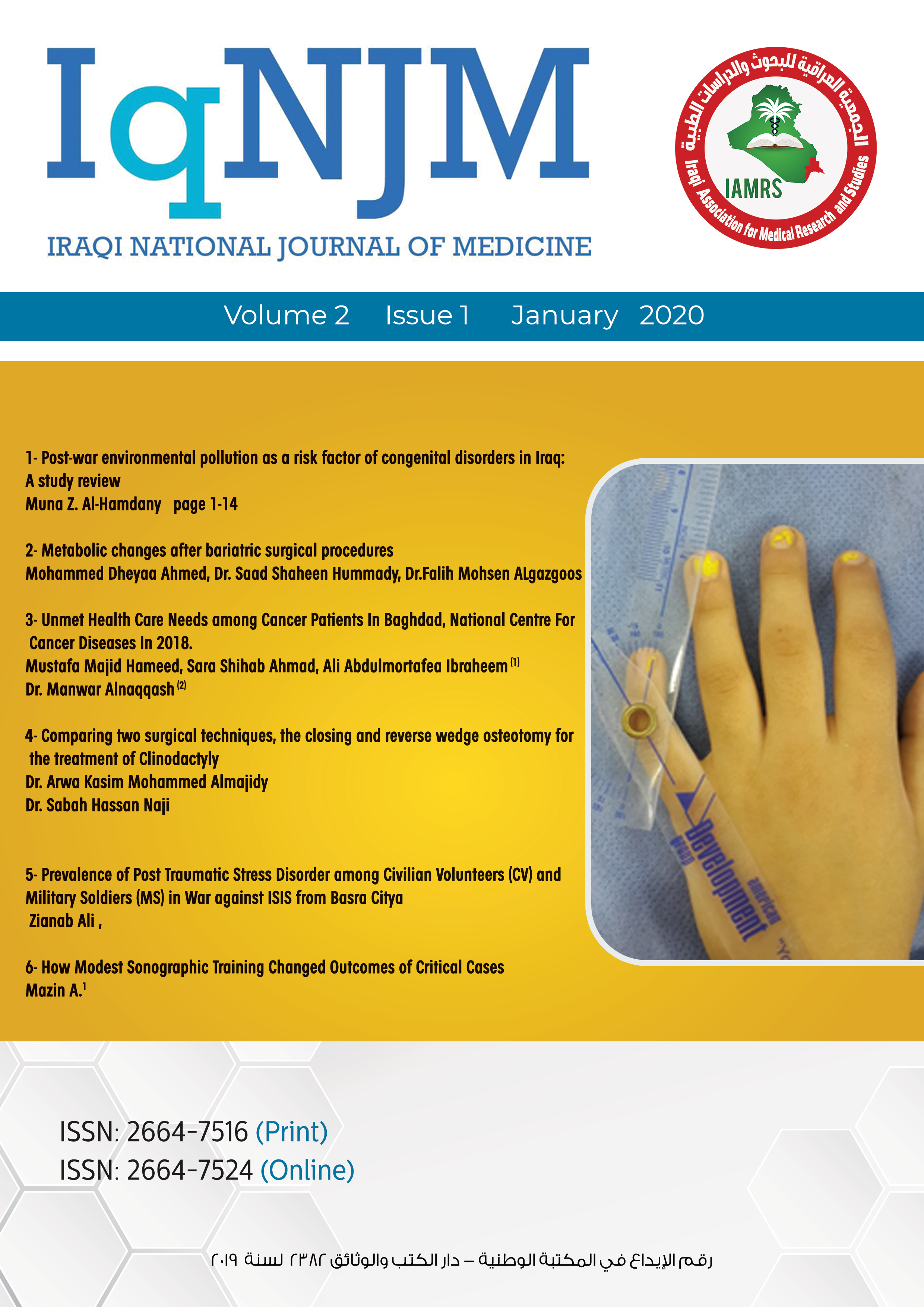
A prospective case study: Comparing two surgical techniques: The closing and reverse wedge osteotomy for treating clinodactyly Closing and reverse wedge osteotomy for the treatment for clinodactyly
Main Article Content
Keywords
clinodactyly, closing wedge osteotomy, reverse wedge osteotomy, congenital hand deformity
Abstract
Background: Clinodactyly or inclination of the digits, particularly the fifth digit, is a congenital anomaly of the hand that occurs in 1% to 19.5% of the population. This deformity requires reconstruction of both the functional and the aesthetic appearance of the finger, if it is severe, to avoid future growth deformity.
Objective: The study aims to review the outcomes and the complications associated with closing and reverse wedge osteotomy techniques for treating clinodactyly.
Patients and Methods: Ten patients’ ten fingers with clinodactyly were submitted for reconstruction from March 2014 to May 2016 in the Al Wasity teaching hospital in Baghdad. They were treated using the closing and reverse wedge osteotomy techniques. In the closing wedge procedure, a wedge was removed from the most convex part of the middle phalanx. Subsequently, the finger is aligned in the midaxial plane and repaired with 2 K-wires. In the reverse wedge osteotomy, the wedge was rotated 180 degrees and reinserted into the bone gap with the wide end first. This buttressed the osteotomy open. Subsequently, the K-wires were inserted in retrograde fashion, maintaining the graft’s position. Then, dressing was applied with the small splint from the PIP to the tip of the finger.
Results: After a 15-month follow-up, all the patients showed satisfactory results aesthetically and the functionally—with full range of motion. There was no recurrence in any case. Only one case had residual angulation and no major complications were encountered.
Conclusion: The closing and reverse wedge osteotomy was proven effective in treating clinodactyly. The closing wedge is simpler than the reverse wedge. The technical difficulty of reverse wedge osteotomy may make it a less appealing option to surgeons but the outcomes we had were rewarding, both techniques provided good overall correction of angulation in one stage, and straightforward procedure, with few complications, good aesthetic outcome and patient satisfaction with improved function.
References
2. Wolfe S, Pederson W, Kozin SH. Part VI. The pediatric hand: Deformities of the hand and fingers. In: Green’s Operative Hand Surgery. 6th ed. Elsevier Health Sciences; 2012. p. 1353.
3. Thorne CH. Part VIII. Hand: Common congenital hand anomalies. In: Grabb and Smith’s Plastic Surgery. 7th ed. 2014. p. 895.
4. Burke F, Flatt A. Clinodactyly. A review of a series of cases. Hand [Internet]. 1979 Oct;11(3):269-80. Available from: http://www.ncbi.nlm.nih.gov/pubmed/520870
5. Evans DM, James NK. A bipedicled neurovascular step-advancement flap for soft tissue lengthening. Br J Plast Surg. 1992;45(5): 380-4.
6. Georgiade GS. Congenital hand deformity. In: FACS Plastic, Maxillofacial and Reconstructive Surgery, Section 8. 3rd ed. 1997. p. 98.
7. Piper SL, Goldfarb CA, Wall LB. Outcomes of opening wedge osteotomy to correct angular deformity in little finger clinodactyly. J Hand Surg Am [Internet]. 2015 May;40(5): 908-913. Available from: https://linkinghub.elsevier.com/retrieve/pii/S0031938416312148
8. Dobyns, Wood, Bayne. Congenital hand deformities. In: Operative Hand Surgery. New York Churchill Livingstone; 1993. p. 426.
9. Ali M, Jackson T, Rayan GM. Closing wedge osteotomy of abnormal middle phalanx for clinodactyly. J Hand Surg Am [Internet]. 2009;34(5): 914-8. Available from: http://dx.doi.org/10.1016/j.jhsa.2009.01.007
10. Carstam N, Theander G. Surgical treatment of clinodactyly caused by longitudinally bracketed diaphysis (“delta phalanx”). Scand J Plast Reconstr Surg. 1975;9(3): 199-202.
11. Al-Qattan MM. Congenital sporadic clinodactyly of the index finger. Ann Plast Surg [Internet]. 2007 Dec;59(6): 682-7. Available from: http://www.ncbi.nlm.nih.gov/pubmed/18046153
12. Light TR, Ogden JA. The longitudinal epiphyseal bracket: Implications for surgical correction. J Pediatr Orthop [Internet]. 1981;1(3): 299-305. Available from: http://europepmc.org/abstract/MED/6801087
13. Gordon AA. Changes in angulation and phalangeal length of fingers and thumbs following surgical treatment for congenital clinodactyly [Internet]. Boston University-School of Medicine; 2014. Available from: https://hdl.handle.net/2144/14696
14. Almajidy AKM. Closing and reverse wedge osteotomy techniques for the treatment of clinodactyly. The Iraqi Board of Medical Specializations in Plastic and Reconstructive Surgery; 2015.


